
What is tissue culture?
Jesus made everything, including plants and their researchers. John 1:3 says all things came into being through Him
(LSB). Regeneration works with capacities He made, under suitable conditions.
Tissue culture is the growth of living cells, tissues, or organs outside the organism under controlled conditions. In plant tissue culture, a small starting piece, called an explant, is placed on or in a nutrient medium and maintained using aseptic techniques. That explant might be a shoot tip, a node carrying a bud, a leaf segment, an embryo, or another plant part chosen for a particular purpose.
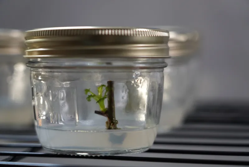
The familiar image is a small green plant inside a clear vessel. Behind it are several connected ideas: supporting living material, managing unwanted microorganisms, supplying suitable nutrition, and understanding how growth changes with the starting tissue and environment. Micropropagation applies these methods to multiplying plants. Tissue culture is the broader field; it also includes experimental systems that do not produce whole plants.
In vitro means that the work takes place outside the organism, commonly in laboratory vessels. It describes the setting, not a promise of success. A culture can be alive without multiplying, and a growing shoot still has a journey before it becomes an established plant. Keeping those distinctions clear makes both historical discoveries and everyday observations easier to understand.
How plant tissue culture works
Living cells need a suitable environment
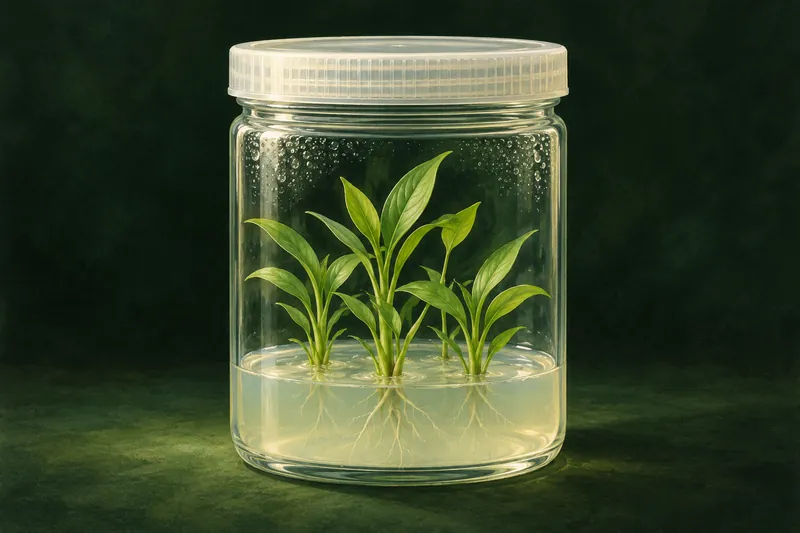
A nutrient medium supports the material being cultured. Depending on the system, it can supply mineral nutrients, vitamins, a carbohydrate source, and plant growth regulators. It may be liquid or supported by a gelling agent. Murashige and Skoog’s influential medium was developed through experiments with tobacco tissue, a reminder that a famous formulation began as a response to a particular biological problem. [11]
The explant matters
“Plant tissue” is not one interchangeable material. A node with an existing bud and a differentiated leaf segment present different starting situations. The plant’s identity, cultivar, age, source conditions, and previous treatment help define what an experiment actually tested. In a study of four blackberry cultivars, researchers evaluated culture establishment, multiplication, and acclimatization within specified treatments; its results belong to those materials and conditions, not every blackberry. [20]
Totipotency is potential, not an automatic outcome
Under suitable conditions, some plant cells can regenerate a whole plant. Scientists investigate this through nutrients, hormones, and cell division. Those explanations describe what Jesus made; they do not replace Him as its Maker.
Totipotency describes the capacity of a cell to generate a complete organism under suitable circumstances. Plant regeneration research made that potential experimentally meaningful. It does not follow that every cell you isolate will regenerate, or that one recipe will make any explant become a plant. The important question is whether the selected material can respond under the conditions provided. Historical experiments progressively tested those conditions rather than assuming the answer.
Clean, sterile, and aseptic mean different things
Cleaning removes soil and residues. Sterilization and disinfection have different purposes and applications; neither should be reduced to “cleaning more.” Aseptic technique concerns how prepared material is handled to avoid introducing unwanted organisms. For practical instruction, use the Academy’s clean-work lessons and a documented procedure appropriate to the materials. This overview explains the field; it is not a substitute for a species-specific protocol.
The history of tissue culture: a shared achievement
Researchers work within Jesus’ creation. Their experiments deserve credit; their lives, materials, and abilities depend on Him. Colossians 1:17 says in Him all things hold together
(LSB; read 1:16–17).
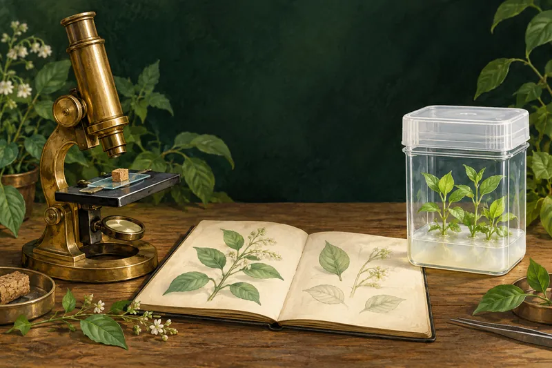
Microscopy, cell theory, plant nutrition, and microbial control laid foundations for later tissue culture. The design came first. The names came later.
1665: Robert Hooke
Hooke described cork compartments as cells in Micrographia. British Library collection account.
1838–1839: Matthias Schleiden and Theodor Schwann
They developed the cellular account of plant and animal structure. Original works in the MBL Rare Books Collection.
1860s: Julius Sachs and Wilhelm Knop
They advanced plant growth in nutrient solutions without soil. Hoagland and Arnon (1950), p. 6.
1860s: Louis Pasteur
Pasteur’s experiments challenged spontaneous generation and connected microbial growth with contamination from the environment. Institut Pasteur historical account.
1867: Joseph Lister
Lister published his antiseptic principle for surgery, an important contribution to controlling contamination in another field. Lister’s original paper.
Late 1800s: Robert Koch
Koch developed bacterial cultivation methods, including solid culture media, that helped researchers isolate and study microorganisms. Koch’s culture-methods paper (1881), RKI archive.
There is no single discovery that explains the entire field. Researchers first had to keep isolated material alive, then demonstrate sustained growth, then understand development and regeneration. Much of that progress depended on collaborators, careful preparation, repeated transfers, and observations that did not initially look like breakthroughs. The milestones below are selected examples, not a complete list of contributors.
1902: Gottlieb Haberlandt asks what isolated plant cells could do
In 1902, Austrian botanist Gottlieb Haberlandt published Culturversuche mit isolierten Pflanzenzellen, describing experiments with isolated plant cells. He did not obtain the cell division needed to establish a continuously growing culture. His lasting contribution was the experimental question: could isolated vegetative cells be supported and eventually develop beyond their existing specialized state? [1]
Haberlandt is often called the father of plant tissue culture. That title recognizes a foundational proposal, not a successful modern cloning protocol. Describing his work this way gives him credit without making later discoveries seem inevitable or suggesting that a missing bottle of hormones alone explains every early difficulty.
1922: Lewis Knudson changes orchid propagation
Lewis Knudson published work on nonsymbiotic orchid seed germination in 1922. He demonstrated an approach to germinating orchid seeds on a prepared medium without the usual fungal association required in their natural setting. It connected nutrition in a vessel with a practical horticultural opportunity. [5]
There is an important distinction here: growing orchid seedlings in culture is not necessarily cloning a selected orchid. Seeds can produce genetically different offspring. Later methods of vegetative propagation addressed a different goal: multiplying a particular plant. Both developments belong in the history, but they should not be described as the same achievement.
1934–1939: Philip White, Roger Gautheret, and Pierre Nobécourt sustain growth
In 1934, Philip R. White reported potentially unlimited growth of excised tomato root tips in liquid medium. Continued growth through transfers helped distinguish a supported culture from a piece of tissue merely surviving for a short time. [2]
By the end of the 1930s, White in the United States and Roger-Jean Gautheret and Pierre Nobécourt in France had established important examples of sustained plant tissue culture. White published on continuing callus growth in 1939; Gautheret and Nobécourt worked with carrot tissues. Their contributions are better credited together than compressed into the name of one inventor. [3] [4]
The practical advance was continuity. A culture could become material for repeated experiments, allowing later researchers to investigate nutrition, growth regulators, and developmental responses. The ability to keep a system going made it possible to ask more precise questions.
1949–1960: Gavino Rotor, Georges Morel, and Claude Martin connect research to propagation
In 1949, Gavino Rotor Jr. described vegetative propagation of Phalaenopsis using flower-stalk material. His work deserves recognition in accounts of orchid propagation that otherwise jump directly from seed germination to later shoot-tip methods. The original description shows how much scientific history can be hidden by an overly short timeline. [6]
In 1952, Georges Morel and Claude Martin reported recovery of dahlias affected by a viral disease using meristem culture. Morel’s later work with orchids helped establish the practical importance of shoot-tip propagation. These contributions linked developmental biology with horticultural production and plant health. [7] [25]
Meristem culture should not be translated into a blanket promise that any plant grown in a jar is virus-free. Plant health claims require suitable starting material, relevant methods, and testing. The achievement was the development of useful approaches to particular problems, not a guarantee attached to every tissue-cultured plant.
1955 and 1957: Carlos Miller, Folke Skoog, and colleagues investigate growth signals
The 1955 kinetin paper credited Carlos O. Miller, Folke Skoog, Malcolm H. von Saltza, and F. M. Strong. Kinetin promoted cell division and became an important early cytokinin. Naming the whole author team gives a more accurate picture than attributing its discovery to only one or two people. [8]
In 1957, Skoog and Miller published their landmark work on chemical regulation of growth and organ formation. Experiments with tobacco tissues showed that changing auxin and cytokinin conditions could change developmental outcomes, including shoot and root formation. [9]
This helped explain why a medium is more than plant food. It can also provide developmental signals. However, the familiar “more cytokinin for shoots, more auxin for roots” explanation is an introduction, not a universal formula. The material and experimental conditions remain part of the result.
1958–1959: Carrot cultures reveal routes to embryo formation
Frederick C. Steward, Marion O. Mapes, and Kathryn Mears published influential studies of growth and organized development in carrot cultures in 1958. Their work belongs to the experimental foundation of somatic embryogenesis: embryo development from somatic, rather than fertilized egg, material. [10]
Jakob Reinert independently investigated embryogenesis in carrot tissue cultures. His widely cited 1959 paper followed observations partly reported in 1958, a chronology he later explained in a first-person account. These parallel lines of research deserve recognition together. [12]
The distinction between making roots or shoots and forming somatic embryos matters. Plants can be regenerated through different developmental routes. A photograph of a green plantlet does not tell the whole story of how that plant was produced.
1962: Toshio Murashige and Folke Skoog publish MS medium
Toshio Murashige and Folke Skoog published their revised medium in 1962. MS medium became a widely adopted starting point for plant tissue culture. Its name preserves both researchers’ contributions. [11]
Today, “MS” can refer to a basal formulation sold in different preparations. A grower still needs to understand what a product contains and what a protocol adds or changes. Basal salts, vitamins, sugar, growth regulators, and gelling agents are not interchangeable categories. Naming a medium is the beginning of documenting a recipe, not the end.
1964–1983: Breeding, protoplasts, and transformation broaden the field
In 1964, Sipra Guha and Satish Chandra Maheshwari reported embryo production from Datura anthers. Anther and pollen culture opened routes relevant to haploid research and plant breeding. Their contribution shows why tissue culture should not be described only as making copies of mature plants. [13]
In 1971, Itaru Takebe, Gudrun Labib, and Georg Melchers reported whole-plant regeneration from isolated tobacco mesophyll protoplasts: cells whose walls had been removed. This provided another striking demonstration of regeneration under suitable conditions. [14]
By 1983, papers such as Robert Fraley and colleagues’ work on bacterial gene expression in plant cells demonstrated important steps in plant genetic transformation. Tissue culture supplied experimental material and regeneration systems for biotechnology; it did not itself mean that a plant had been genetically engineered. Many ordinary micropropagation projects involve no introduced genes at all. [15]
The parallel history of animal and human tissue culture
Plant tissue culture developed alongside a broader field. Ross Granville Harrison’s pioneering experiments, associated with 1907, made growing nerve fibers from embryonic frog tissue observable outside the animal. Yale’s archival account places this work among the foundations of experimental tissue culture. [16]
In 1951, cells taken from Henrietta Lacks without her knowledge or consent became the source of the HeLa cell line. Her contribution must be acknowledged alongside the laboratory work. George Gey’s team established cultures that became widely used in biomedical research. NIH’s account explicitly recognizes Lacks, her family, and the circumstances in which the cells were obtained. Scientific usefulness does not remove the importance of consent and respectful attribution. [17]
John Enders, Thomas Weller, and Frederick Robbins received the 1954 Nobel Prize for showing that poliovirus could grow in cultures of different tissues. Their work helped enable subsequent vaccine development. This medical branch shares the challenge of supporting living material outside an organism, while its methods and purposes differ substantially from propagating a blackberry or orchid. [18]
The plant tissue culture journey: from explant to established plant
The stages work with the plant’s growth and development—the design Jesus made. Learn the sequence before starting: prepare materials and follow the chosen protocol’s timing and handling.
For growers, the history becomes practical through a sequence of work. The stages below describe a common micropropagation pathway; laboratories may divide or name them differently. Embryo culture, conservation, breeding, and cell-production systems can follow other routes. FAO’s propagation guidance illustrates how culture establishment, multiplication, rooting, and acclimatization connect in an actual crop system. [19]
Choose a stage to explore its purpose, the questions to ask, and what to record.
1. Source
Select and document the source. Record the plant, cultivar if known, donor condition, and intended explant. Decide what result the protocol is designed to produce.
- The aim
- Start with an identity you can follow.
- A question to ask
- Which plant and which starting tissue are involved?
- Keep in the record
- Plant identity, cultivar if known, donor condition, explant type.
2. Initiation
Establish the culture. Introduce prepared material into suitable conditions. Initiation concerns establishing useful living cultures, not simply placing tissue in a vessel.
- The aim
- Establish useful living cultures.
- A question to ask
- What changed after the explant entered culture?
- Keep in the record
- Introduction date, initial appearance, observations and next check.
3. Multiplication
Multiply or develop the material. Follow the chosen propagation route and document each transfer. Record useful growth rather than relying only on how full a vessel looks.
- The aim
- Follow useful growth through each transfer.
- A question to ask
- What can actually continue into the next cycle?
- Keep in the record
- Parent batch, transfer date, useful shoots and the protocol version.
4. Development
Prepare for independent growth. Rooting and plantlet development must be considered in relation to the next stage, not treated as isolated photographs of success.
- The aim
- Prepare plantlets for their next environment.
- A question to ask
- What does the selected protocol require before transfer?
- Keep in the record
- Rooting or development observations and readiness criteria.
5. Acclimatization
Acclimatize and evaluate. Follow the plant beyond the vessel. Record survival and subsequent growth so the result includes the transition to its next environment.
- The aim
- Follow the plant beyond its vessel.
- A question to ask
- Does establishment continue after transfer?
- Keep in the record
- Transfer conditions, survival, subsequent growth and outcome.
A useful notebook can ask the same questions at every stage: What did I start with? What conditions did I provide? What changed? What did I do next? What happened afterward? Those questions help a grower learn from both strong and disappointing outcomes. They also make comparisons more meaningful when a recipe, supplier, cultivar, or handling step changes.
What tissue culture makes possible—and what it does not guarantee
Propagation and conservation
Micropropagation can support multiplication of selected plant material, while in vitro collections and cryopreservation can contribute to conservation. FAO’s genebank standards treat these as managed systems with requirements for documentation, viability, security, and distribution. A conserved accession is valuable because its identity and condition remain traceable, not merely because tissue is stored somewhere. [21]
For a small grower, that suggests a practical discipline: connect the parent material to each later batch. For a research collection, it means keeping identity and provenance attached to the material through storage and recovery. Scale changes the logistics, but losing the identity weakens the meaning of the result.
Clonal propagation does not mean variation is impossible
In 1981, Peter Larkin and William Scowcroft drew attention to somaclonal variation: variability associated with plants regenerated from cultured material. Their work helped make clear that tissue culture can be a source of variation as well as a tool for multiplication. Uniformity must therefore be evaluated rather than assumed. [22]
A successful protocol has boundaries
A published result should travel with its plant material, growth conditions, measurements, and limitations. “It worked in one experiment” is useful evidence, but it is not the same statement as “it will work in every cultivar.” Before adopting a method, look for the actual species, explant, stage, treatment, and reported outcome. If those details do not match your situation, label the method as something to evaluate.
What should you watch for in your own cultures?
A culture offers observations before it offers explanations. Useful records describe where a change appeared, when it began, and whether growth continued. “Brown area at the cut, shoot still growing” is more informative than “bad jar.” It leaves room to investigate the cause without rewriting what was actually seen.
Research on Medinilla formosana documented browning responses after cutting and examined associated changes in gene expression. It supports treating cutting-related browning as a biological response worth investigating, but it does not diagnose a blackberry culture. A localized brown wound and a declining shoot are different observations, and neither should be converted automatically into a confirmed contaminant name. [23]
Use a photograph through the closed vessel when possible, date the observation, and compare it with later development. Keep four separate notes: observation, interpretation, action, and outcome. If your interpretation changes, preserve the earlier observation. That creates a learning record instead of a story rewritten to fit the ending.
MyTCLab connects preparation, batches, locations, and journal records so those details can stay together. The software cannot turn an unsupported explanation into a validated protocol. Its purpose is to make the work easier to organize and the evidence easier to revisit.
Common questions about tissue culture
Is tissue culture the same as micropropagation?
Micropropagation is an application of plant tissue culture aimed at multiplying plants. The wider field also includes cell cultures, organ cultures, experimental regeneration, breeding methods, and conservation systems.
Are tissue-cultured plants genetically modified?
The propagation method alone does not establish that. Ordinary clonal multiplication and genetic transformation are different activities, even though transformation research may use tissue culture and regeneration techniques.
Does growing in a sterile vessel guarantee a disease-free plant?
No. A vessel’s appearance and a plant’s tested health status are different kinds of evidence. Claims about freedom from particular pathogens need an appropriate production and testing process.
Can beginners learn plant tissue culture?
Yes. Start by understanding the workflow, measurement, clean handling, and the purpose of each stage. Choose a documented starting method, keep the scope manageable, and judge progress through records rather than expecting every vessel to succeed.
Keeping the knowledge accessible
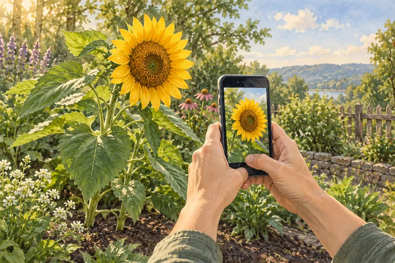
People respond with hearts and likes to a beautiful sunflower photograph on social media. Jesus made the sunflower, the photographer, and the world around them. Our life, breath, will, and ability to create depend on His sustaining care. Becoming aware means recognizing Him throughout the scene and giving Him thanks.
The field reached today’s growers through more than landmark papers. Technicians, teaching laboratories, commercial propagators, botanical collections, and patient educators made techniques repeatable and understandable. A fair account recognizes that contribution without pretending this selected history names everyone who deserves credit.
Modern educators also help people take a first step. Laur of Plants in Jars explains tissue culture through accessible tutorials, supplies, and practical demonstrations. Laur’s own introduction describes beginning as a hobbyist and creating the straightforward explanations that were difficult to find. [24]
For MyTCLab founder Steward Clinton, Laur’s teaching helped demystify the process and inspired the move from curiosity to hands-on learning. That is a personal acknowledgment of influence, not a claim that Laur invented tissue culture or endorses MyTCLab. Helping another person understand what is possible is a meaningful contribution to participation in the field.
If this overview has made the subject clearer, continue with the free Tissue Culture Academy. It covers plant context, clean workflow, media, measurement, culture stages, troubleshooting, and evidence. The next useful step is not memorizing every date. It is understanding the next part of your work well enough to carry it out thoughtfully, observe the result, and learn from it.
Scripture: Legacy Standard Bible (LSB)
Scripture names the Creator; research documents observations. Short LSB excerpts link to full passages.
- John 1:3 (LSB)
- Colossians 1:16–17 (LSB)
- Hebrews 1:2 (LSB):
He made the worlds
. - Genesis 1:11–12 (LSB):
plants yielding seed
. - Romans 1:20 (LSB):
being understood through what has been made
.
References and original publications
- Haberlandt, G. (1902). Culturversuche mit isolierten Pflanzenzellen. Original publication, Biodiversity Heritage Library. Read source 1
- White, P. R. (1934). Potentially Unlimited Growth of Excised Tomato Root Tips in a Liquid Medium. Plant Physiology 9:585–600. Read source 2
- White, P. R. (1939). Potentially Unlimited Growth of Excised Plant Callus in an Artificial Nutrient. American Journal of Botany. Read source 3
- Nobécourt, P. (1942). Sur la culture illimitée des tissus végétaux. First-person account discussing the early carrot cultures and Gautheret’s parallel work. Read source 4
- Knudson, L. (1922). Nonsymbiotic Germination of Orchid Seeds. Botanical Gazette 73:1–25. Read source 5
- Rotor, G. Jr. Vegetative Propagation of Phalaenopsis. Original article scan hosted by North Carolina State University; related 1949 account in the American Orchid Society archive. Read source 6
- Morel, G., and Martin, C. (1952). Guérison de dahlias atteints d’une maladie à virus. Comptes rendus de l’Académie des sciences 235:1324–1325. Read source 7
- Miller, C. O., Skoog, F., von Saltza, M. H., and Strong, F. M. (1955). Kinetin, a Cell Division Factor from Deoxyribonucleic Acid. JACS 77:1392. Read source 8
- Skoog, F., and Miller, C. O. (1957). Chemical regulation of growth and organ formation in plant tissues cultured in vitro. Symposia of the Society for Experimental Biology 11:118–130. Read source 9
- Steward, F. C., Mapes, M. O., and Mears, K. (1958). Growth and Organized Development of Cultured Cells. II. Organization in Cultures Grown from Freely Suspended Cells. American Journal of Botany 45:705–708. Read source 10
- Murashige, T., and Skoog, F. (1962). A Revised Medium for Rapid Growth and Bio Assays with Tobacco Tissue Cultures. Physiologia Plantarum 15:473–497. Read source 11
- Reinert, J. (1984). First-person commentary on his 1959 carrot embryogenesis paper and observations reported in 1958. Citation Classics, University of Pennsylvania archive. Read source 12
- Guha, S., and Maheshwari, S. C. (1964). In vitro Production of Embryos from Anthers of Datura. Nature 204:497. Read source 13
- Takebe, I., Labib, G., and Melchers, G. (1971). Regeneration of whole plants from isolated mesophyll protoplasts of tobacco. Naturwissenschaften 58:318–320. Read source 14
- Fraley, R. T., and colleagues (1983). Expression of bacterial genes in plant cells. PNAS 80:4803–4807. Read source 15
- Yale School of Medicine. History of the Department of Cell Biology (2011), including Harrison’s early tissue-culture experiments. Read source 16
- National Institutes of Health (2013). Guidance on acknowledgement and use of HeLa cell whole-genome sequence data, recognizing Henrietta Lacks and her family. Read source 17
- Enders, J. F., Robbins, F. C., and Weller, T. H. (1954). The Cultivation of the Poliomyelitis Viruses in Tissue Culture. Nobel Lecture. Read source 18
- Zaid, A., and de Wet, P. F. Date Palm Propagation. FAO, chapter V. Crop-specific guidance, not a universal protocol. Read source 19
- Enhancing In Vitro Multiplication and Acclimatization of Blackberry (Rubus L.) Through Sterilization Optimizing and Growth Regulator Use (2025). Horticulturae 11:1422. Four cultivars studied; not Ouachita validation. Read source 20
- FAO. Genebank Standards for Plant Genetic Resources for Food and Agriculture: in vitro culture and cryopreservation. Read source 21
- Larkin, P. J., and Scowcroft, W. R. (1981). Somaclonal variation—a novel source of variability from cell cultures for plant improvement. Theoretical and Applied Genetics 60:197–214. Read source 22
- Characterization and Comparative Expression Profiling of Browning Response in Medinilla formosana after Cutting (2016). Original research; species-specific evidence. Read source 23
- Laur, Plants in Jars. About Plants in Jars and beginner tutorial information. First-person account; checked October 17, 2026. Read source 24
- Morel, G. (1960). Producing Virus-Free Cymbidiums. American Orchid Society Bulletin 29:495–497; original July 1960 issue archive. Read source 25